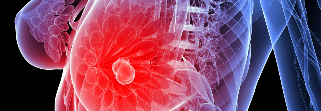
3D Mammography – Digital Tomosynthesis

Digital tomosynthesis (pronounced toh-moh-SIN-thah-sis), creates a 3-dimensional picture of the breast using X-rays. 3D Mammography (digital tomosynthesis), is approved by the U.S. Food and Drug Administration but is not yet considered the standard of care for breast cancer screening. It is a very new technology and is available at Thousand Oaks Radiology.
What is the difference between Digital Mammography and 3D Mammorgraphy?
Digital mammography usually takes two X-rays of each breast from different angles: top to bottom and side to side. The breast is pulled away from the body, compressed, and held between two glass plates to ensure that the whole breast is viewed. Digital mammography records the pictures on the computer. The images are then read by our radiologist. Breast cancer, which is denser than most healthy nearby breast tissue, appears as irregular white areas — sometimes called shadows.
3D Mammography (Digital tomosynthesis), is a new kind of test that takes multiple X-ray pictures of each breast from many angles. The breast is positioned the same way it is in a conventional mammogram. The X-ray tube moves in an arc around the breast while 9 images are taken during the examination. Then the information is sent to a computer, where it is assembled to produce clear, highly focused 3-dimensional images throughout the breast.
Breast tomosynthesis may also result in:
- fewer unnecessary biopsies or additional tests.
- greater likelihood of detecting multiple breast tumors.
- clearer images of abnormalities within dense breast tissue.
- greater accuracy in identifying the size, shape and location of breast abnormalities.
- earlier detection of small breast cancers that may not be detected on a conventional mammogram.
Early results with 3D Mammography (digital tomosynthesis) are promising. At Thousand Oaks Radiology, we believe that this new breast imaging technique will make breast cancer easier to detect in patients with dense breasts.


For more information on preparation, see Digital Mammography.

